Related field
AI scientific illustration in 2026: figures for papers, posters and grants that don't look generated
Updated 1 June 2026 · 11 min read

AI scientific illustration is, in 2026, useful for schematics, conceptual diagrams and figure polish, and unsafe for anything that depends on factual structure the model has to invent. This guide covers which figure types AI genuinely speeds up, the journal disclosure rules now in force, a reproducible sketch-first workflow, and the small list of tools worth knowing for papers, posters and grants.
What 'AI scientific illustration' actually means in 2026
AI scientific illustration is the use of generative models to draft, render or annotate figures for research output: papers, posters, grants, theses and talks. The useful definition narrows that further. A figure has to be correct, original and reproducible. Text-to-image generation fails all three: it averages training data of mixed quality, produces stochastic outputs that you cannot regenerate identically, and learns from copyrighted figures with fuzzy provenance. Sketch-first and AI-assisted refinement workflows preserve all three properties because the scientific content originates with the author.
The other framing that matters is who the figure is for. A figure for an internal lab meeting tolerates more AI involvement than one going to Nature. The model's job, in any of these settings, is the same: take a structurally correct input and improve the rendering, layout, labelling or colour. It is never to decide what a pathway looks like, where an arrow goes, or which residues sit in an active site.
Why generic image models fail scientific figures
Models like DALL-E 3, Midjourney v7 and Stable Diffusion XL were trained on billions of web images. The web contains a vast amount of mis-labelled or simplified scientific imagery, AI-recycled figures, and species-mixed anatomy. The model averages all of it and rewards plausibility, not accuracy. A 2024 npj Digital Medicine paper testing four leading text-to-image models on standard anatomy prompts found anatomically faithful outputs in under 15% of cases. For molecular structures the failure rate is higher still: stereochemistry, ring saturation and bond orders are routinely wrong.
There is a second problem that matters more for science than for medicine: reproducibility. The same prompt to the same model on the same day will not produce the same image, and the seed control most image APIs expose is unreliable across model updates. A figure you cannot regenerate from a published prompt is a figure a reviewer cannot audit. That alone disqualifies pure text-to-image for any methods-grade figure.
Which figure types AI scientific illustration genuinely helps with
Across two years of regular use in academic labs, a stable shortlist has emerged. AI accelerates schematics, conceptual diagrams, draft posters and figure polish. It does not help, and often hurts, on data plots, histology, microscopy, structural biology and molecular structures, where mature non-AI tools already exist and produce ground-truth output. The table below maps figure types to the tool class that earns its place.
| Figure type | Best tool class (2026) | Why | Avoid |
|---|---|---|---|
| Pathway / signalling diagrams | BioRender, Mind the Graph | Curated icon libraries, conventional aesthetic | Text-to-image |
| Experimental schematics | Sketch-first AI (Angiosome) | You set structure, AI handles render | Generic Midjourney prompts |
| Conceptual / mechanistic figures | Sketch-first AI or Inkscape + SciDraw | Originality, accurate to your model | Text-to-image |
| Anatomical figures | Sketch-first AI for diagrams; real images for histology | Anatomy must be controlled; histology must be real | DALL-E, Midjourney histology |
| Molecular structures | ChemDraw, RDKit | Stereochemistry, bond orders correct by construction | Any image AI |
| Structural biology | PyMOL, ChimeraX | Generated from coordinate files; reproducible | Any image AI |
| Data plots | ggplot2, matplotlib, Plotly | Not an AI problem; reproducibility from data | AI redraws of charts |
| Posters | Canva or LaTeX layout + AI-rendered panels | Layout conventional; panels can be AI-assisted | Single text prompt for whole poster |
Journal policy on AI figures: what 2026 actually requires
Most major journals updated their AI policies between 2023 and 2025 and the consensus has firmed up. The ICMJE 2023 recommendations require authors to disclose any generative AI used in figures and to take full responsibility for accuracy. Nature does not currently accept fully AI-generated figures in submitted manuscripts but allows AI-assisted refinement of human-drawn artwork. JAMA and the BMJ allow disclosed AI use. Science and Cell broadly mirror the Nature position. Assume disclosure is required unless the journal explicitly says otherwise, and check the target journal's instructions for authors before submission.
| Publisher | Fully AI-generated figures | AI-assisted refinement | Disclosure required |
|---|---|---|---|
| Nature | Not accepted | Allowed with disclosure | Yes |
| Science | Restricted | Allowed with disclosure | Yes |
| Cell Press | Restricted | Allowed with disclosure | Yes |
| JAMA Network | Allowed with disclosure | Allowed with disclosure | Yes |
| BMJ | Allowed with disclosure | Allowed with disclosure | Yes |
| NEJM | Restricted | Allowed with disclosure | Yes |
| PLOS | Allowed with disclosure | Allowed with disclosure | Yes |
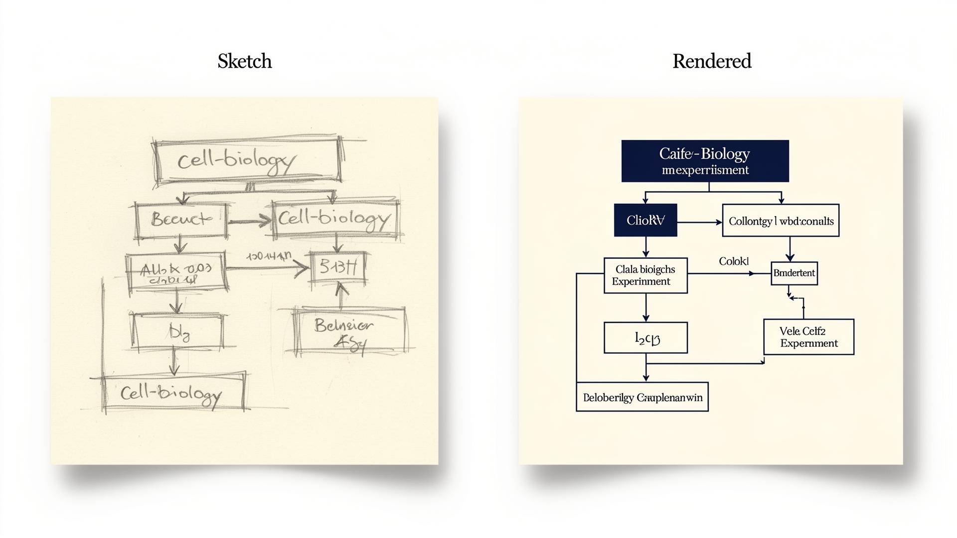
A reproducible workflow for AI-assisted scientific figures
This is the workflow used most often by labs that have settled on AI as part of their figure pipeline rather than a one-off experiment. It assumes one figure for a paper or grant. Total time per figure once you've used it a few times: around 30 minutes for a schematic, longer for a multi-panel figure.
- Sketch the figure on paper or a tablet. Mark every structure, label, arrow and panel boundary. Treat this as the scientific content of the figure.
- Render the schematic with a sketch-first AI tool (Angiosome or similar) or assemble it from a vector library (BioRender, Mind the Graph, SciDraw). Aim for two iterations: rough render, then refined with corrected labels.
- Compose the final figure in your vector tool of choice (Illustrator, Inkscape, Figma). Add typography, scale bars, panel letters and any data overlays.
- Export at journal resolution: 300 dpi minimum for raster, native SVG/EPS where the journal accepts vector. Most journals now specify 300 dpi for line art and 600 dpi for combined figures.
- Archive provenance: source sketch, tool name and version, prompts used, intermediate renders. Add a one-line disclosure to the figure legend or methods section: 'Figure X was drafted using [tool, version]. Final composition by [author]. All scientific content verified by the authors.'
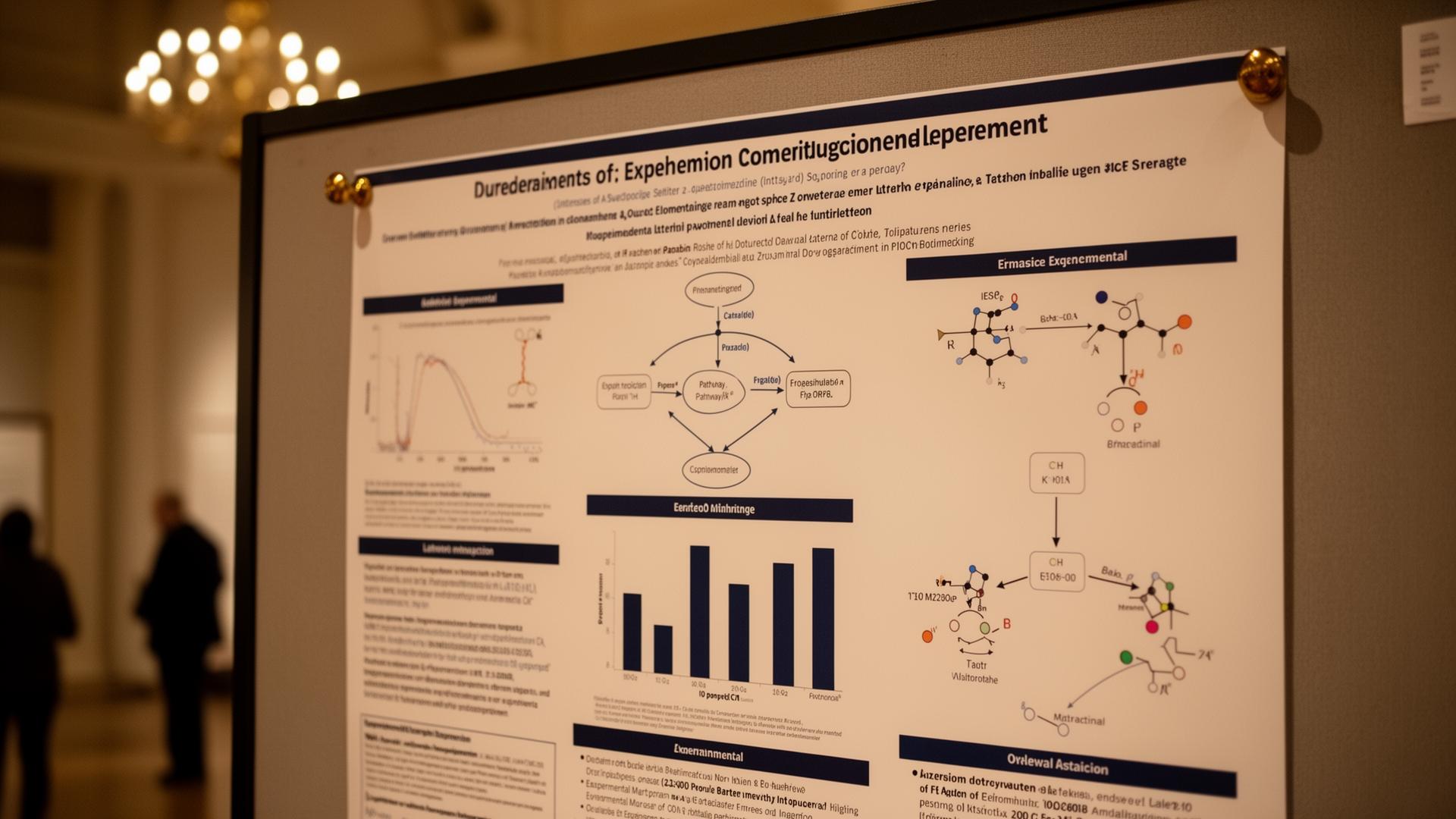
Posters and grants: where AI saves the most time
Conference posters and grant figures are the highest-leverage use case for AI scientific illustration because the audience is generous about aesthetic and the figures are usually conceptual, not methods-grade. A typical poster needs an overview schematic, a results panel and a model figure. AI can draft the schematic and the model figure in under an hour combined; the results panel comes from your data and is not an AI problem. Grant figures are similar: a methods overview and a conceptual model, both of which an AI-assisted workflow handles well.
BioRender and the close alternatives, ranked honestly
BioRender remains the default for biological pathway figures because its icon library is the largest in the category (around 50,000 icons across 30+ disciplines in 2026) and the drag-and-drop workflow is genuinely fast. Its weaknesses are price (Premium $39/month billed annually, Pro $79/month), a free tier that explicitly bans publication use, and a house style instantly recognisable to reviewers. Mind the Graph is the closest like-for-like competitor at $10–$25/month with attribution-permitted free publication. Inkscape combined with SciDraw and Reactome icons reaches publication quality at zero recurring cost but takes roughly 3x longer per figure while you learn the vector workflow. The deeper comparison is in the biorender-alternatives guide.
What AI scientific illustration still does badly
- Histology and microscopy. Use real images from your lab, Pathology Outlines, or open repositories. Never let AI generate tissue.
- Molecular structures with stereochemistry, ring saturation or bond orders. Use ChemDraw or RDKit-rendered structures from SMILES.
- Crystallography and structural biology. Render from coordinate files in PyMOL or ChimeraX. The figures are reproducible from the PDB ID alone.
- Reproducing a specific published figure's style as your own. Both an originality and a copyright problem; don't.
- Generating data plots from numbers. Use ggplot2, matplotlib, or Plotly. The plot script is part of your reproducibility record.
- Anything where a small visual error changes the scientific claim (active-site geometry, gating positions, electrophysiology traces). The category of figure where 'plausible-looking' is most dangerous.
Sources
- ICMJE Recommendations on the use of generative AI in scholarly publishing — ICMJE, 2023
- Nature editorial policy on generative AI in figures and text — Nature
- JAMA Network instructions for authors on AI use — JAMA Network
- BMJ author guidance on generative AI — BMJ
- BioRender — official pricing page — BioRender
- SciDraw — open scientific drawings — SciDraw
- Reactome — pathway database and icons — Reactome
- Inkscape — open-source vector editor — Inkscape Project
Frequently asked questions
Can I use AI to make figures for a scientific paper?
Yes, with disclosure and within the target journal's policy. AI is safe for schematics, conceptual diagrams and figure polish where you supply the structure and the model only renders. Avoid AI for histology, microscopy, molecular structures and crystallography because the failure modes are scientific, not cosmetic. Keep your source sketches, prompts and intermediate renders as part of the figure provenance record.
What is the best AI tool for scientific illustration in 2026?
It depends on figure type. BioRender or Mind the Graph for pathway and cell biology figures; sketch-first AI tools like Angiosome for experimental schematics, anatomy and conceptual diagrams; Inkscape with SciDraw and Reactome icons for full editorial control at zero cost; ChemDraw, PyMOL and ChimeraX for molecules and structures. No single tool wins across all figure types.
Do journals require disclosure of AI use in figures?
Yes, in nearly all major biomedical and life-science journals. The ICMJE 2023 update requires authors to disclose any generative AI used in figures and take full responsibility for accuracy. Nature, Science, Cell, NEJM, JAMA, BMJ and PLOS all enforce this. A single line in the figure legend or methods section is usually enough; always check the target journal's specific instructions before submission.
Is AI scientific illustration accepted by Nature and Science?
Refinement is, fully generated figures generally are not. Nature does not accept fully AI-generated figures in submitted manuscripts but allows AI-assisted refinement of human-drawn artwork with disclosure. Science and Cell take a similar position. JAMA and BMJ allow disclosed AI-generated figures. The safe default for top-tier journals is sketch-first or vector-library workflows where the scientific content originates with the author.
How does Angiosome compare to BioRender for scientific figures?
Angiosome and BioRender solve different problems. BioRender wins on standardised pathway and cell biology figures because its icon library is the largest in the category. Angiosome wins on experimental schematics, anatomy, and any structure where you need a sketch-first workflow because the model preserves your topology instead of inventing one. Most labs use both, picking by figure type rather than committing to one platform.
Can AI generate molecular structures or crystal structures?
No, not reliably. Generative image models routinely produce wrong stereochemistry, impossible ring systems and invalid bond orders. Use ChemDraw or RDKit rendered from SMILES strings for chemistry, and PyMOL or ChimeraX rendered from coordinate files for structural biology. These tools generate from ground truth and are reproducible from a single identifier, which is what reviewers and editors expect.
Will using AI for figures get my paper rejected?
Disclosed, accurate, AI-assisted figures are increasingly accepted across biomedical publishing. Undisclosed fully AI-generated figures, particularly at top-tier journals like Nature, Science and Cell, are increasingly rejected. The pattern that survives peer review: sketch-first or vector-library workflows, transparent disclosure in the methods section, and retained provenance for any reviewer who asks. Treat AI as a drafting tool, not an unattributed co-author.
What is the cheapest way to produce publication-quality scientific figures?
Inkscape combined with SciDraw and Reactome icon libraries is fully free, produces journal-grade SVG, and satisfies CC-BY attribution requirements. The trade-off is time: budget roughly three times longer per figure than a drag-and-drop tool while you learn Inkscape's layer and path model. For one-off figures most researchers find this worthwhile; for high figure volume, paying for BioRender or Mind the Graph saves more time than it costs.
Try it
Sketch it. Angiosome renders it.
Angiosome turns rough medical sketches into clean, labelled, photoreal diagrams — grounded in your sketch, not invented by a model. Free to try.
Open Angiosome →